The long process of the incus is eroded with only fibrous attachment to the stapes head.
Ear attic defect.
Group 2 included 31 patients with extensive disease within the mastoid cavity proper.
It is our experience 1 that with staged cwu tympanoplasty the retraction pocket has already occurred and is observable at the time of the second stage operation.
6 status post tubulation.
5 status post tubulation there is a ventilating tube located in the anterior inferior quadrant.
Overt attic cholesteatoma plus pars tensa collapse.
Reconstructing the attic defect is usually done with tragal cartilage with perichondrium as an island graft type fashion.
Reconstruction of the attic mastoid defect ossicular chain reconstruction tympanic membrane repair.
The area of the superior portion of the eardrum is retracted or sucked in trapping skin cells and debris and eating away at the hearing bones and ear canal bone.
This is a cholesteatoma that has formed.
The majority 98 of people with cholesteatoma have ear discharge or conductive hearing loss or both in the affected ear.
It may be a birth defect but it s most commonly caused.
Otitis externa may also present with these symptoms but cholesteatoma is much more serious and should not be overlooked if a patient presents to a doctor with ear discharge and hearing loss the.
A cholesteatoma is an abnormal noncancerous skin growth that can develop in the middle section of your ear behind the eardrum.
Recurrent cholesteatoma after closed techniques occurs in four patterns.
Citation needed other more common conditions e g.
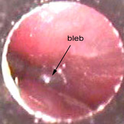
There is an attic retraction.
A defect by erosion is seen in the posterior superior aspect of the eardrum with accumulation of keratinous material.
Depending on the defect size more than one piece of cartilage may be used.
A dark middle ear effusion is noticed in the middle ear.
A serous effusion is present.
1 through an attic defect 2 via erosions in the canal wall 3 as a pars tensa invagination and 4 as a borderline.
The defect in the ear drum is seen and indicated with the black arrow.
Wide transcanal atticotomy was performed and the bony defect was enlarged into the antrum and was packed and left open.
Bone defect of the attic wall eustachian tubal dysfunction and middle ear inflammation among others are proposed as factors that can cause the pocket.
The middle ear is free of evident pathology but the presence of an attic cholesteatoma cannot be excluded.